Kidney Cancer
Robotic Surgery for Kidney Cancer: Partial & Radical Nephrectomy
A complete guide to robotic kidney cancer surgery — single-port retroperitoneal partial nephrectomy through a small low incision, radical nephrectomy when the kidney can't be saved, and what recovery genuinely looks like.
Most kidney tumors today are found by accident — a CT or ultrasound ordered for something else entirely turns up a mass, and suddenly you’re reading pages like this one. Two things are worth knowing up front: most localized kidney cancers are highly curable with surgery, and in most cases the operation can save the rest of your kidney. This page explains how I approach kidney masses, how the two operations work — partial and radical nephrectomy — and what recovery genuinely looks like.
First: not every kidney mass needs surgery tomorrow
An honest starting point matters here. Most solid kidney masses are cancerous, and many grow quickly — a kidney mass should never be ignored or put off. But treating it seriously doesn’t automatically mean surgery for everyone: depending on your age, overall health, and the size and behavior of the mass, the right plan may be active surveillance (close monitoring with periodic imaging, reserved for carefully selected patients), ablation (destroying a small tumor with heat or cold), or surgery. I walk every patient through this menu honestly — surgery is what I do, but it’s only what I recommend when it’s truly the right tool for your situation.
When surgery is the right tool, the next question is: how much kidney has to go?
Partial vs. radical: saving the kidney comes first
Your kidneys filter your entire blood volume many times a day — every ounce of working kidney tissue you keep is filtration capacity you keep for life.
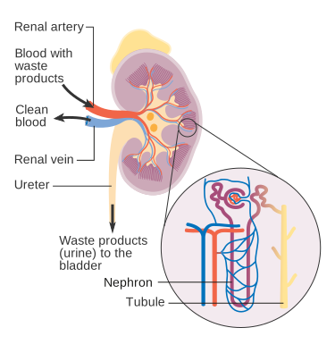
The kidney filters blood through millions of microscopic nephrons — tissue worth preserving. Illustration: Cancer Research UK, CC BY-SA 4.0

- Partial nephrectomy removes the tumor with a rim of healthy tissue and reconstructs the kidney, preserving the rest of it. This is my goal whenever the tumor’s size and location allow. Keeping kidney tissue matters — for your long-term kidney function, your blood pressure, and your options if anything ever happens to the other kidney.
- Radical nephrectomy removes the entire kidney. It’s the right operation when the tumor is large, sits centrally where reconstruction isn’t feasible, or has replaced too much of the kidney to save.
Cancer control is equivalent when each operation is chosen appropriately — this isn’t a trade-off between cure and kidney preservation. It’s about not removing more of you than the cancer requires.
My signature approach: single-port through a low anterior incision
One of my specialties is single-port retroperitoneal partial nephrectomy through a low anterior access incision — an approach I use as my default, and one that few centers in the Pacific Northwest offer.
Here’s what makes it different. The kidney sits behind the abdominal cavity, in a space called the retroperitoneum. Most robotic kidney surgery in the U.S. reaches it the long way: through the front of the abdomen with five or six incisions, entering the space where your intestines live and moving them aside. The traditional alternative — flank retroperitoneal access — avoids the abdomen but requires positioning you on your side and struggles to reach tumors on the front of the kidney.
The low anterior access changes the geometry: through one small incision (about 4 cm) low on the abdomen, near the beltline, I enter the retroperitoneal space directly and bring the single-port robot to the kidney from below. You lie flat on the table — no flank positioning — and the peritoneal cavity is never opened.
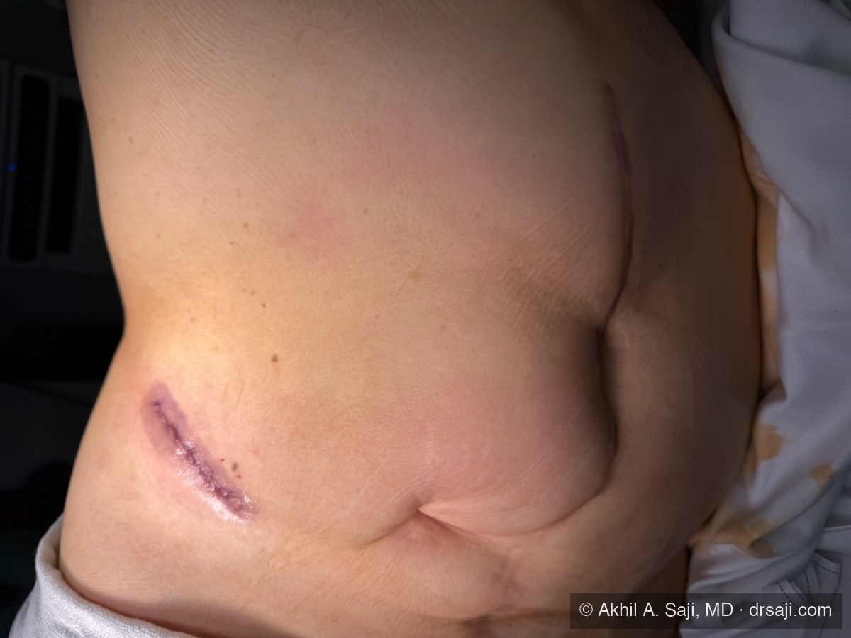
Day of surgery
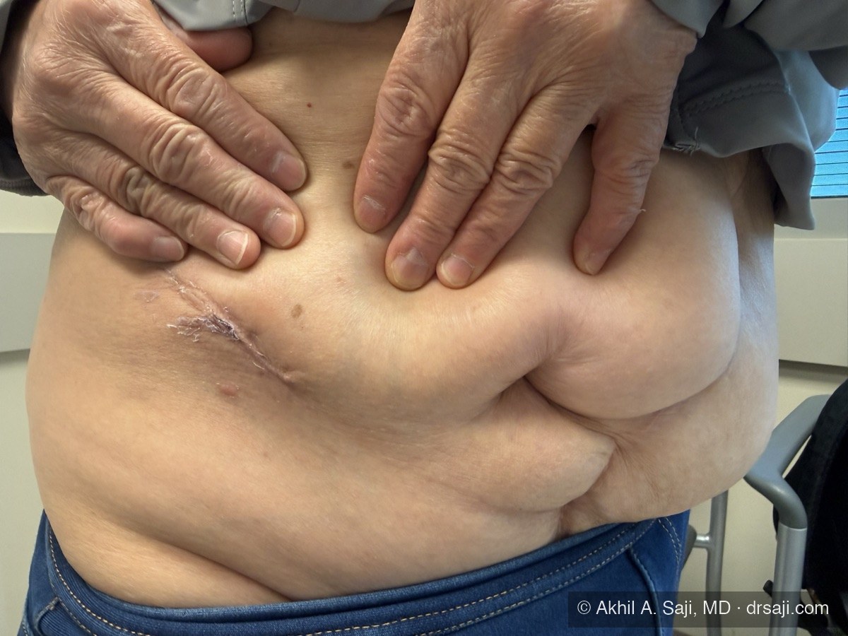
Day 10 follow-up
Why this matters to you:
- Your bowels are never touched. In a large multicenter comparison against standard multiport surgery, this approach cut opioid use nearly in half and eliminated post-operative bowel shutdown — the single most miserable part of traditional abdominal surgery.
- Any tumor location. Unlike classic flank retroperitoneal surgery, the low anterior access reaches tumors on the front, back, and side of the kidney.
- Prior surgery is not a barrier. If you’ve had abdominal operations before — hernia repairs with mesh, bowel surgery, a C-section — the scar tissue lives in the space I never enter. Patients told elsewhere that their abdomen is too “hostile” for minimally invasive surgery are often straightforward candidates this way.
- One small scar near the beltline, not a constellation across your abdomen.
One important caveat: single-port is not the right tool for every kidney. For some tumors — depending on size, complexity, location, and your individual anatomy — classic flank retroperitoneal access or traditional multiport robotic surgery remains the better and safer choice, and I use those approaches regularly for exactly that reason. The approach serves the operation, not the other way around. Which one is right for you is a decision we make together at your consultation, after I’ve reviewed your imaging.
How partial nephrectomy works, step by step
- Anesthesia and positioning. You’re fully asleep, lying flat.
- Access. One small incision low on the abdomen; I develop the space behind the peritoneum and dock the SP robot.
- Exposure. I dissect directly onto the kidney and its blood vessels and locate the tumor, guided by your imaging and an ultrasound probe on the kidney itself.
- Tumor removal. Blood flow to the kidney is briefly paused so I can remove the tumor cleanly with a margin of healthy tissue. I keep that pause as short as possible — and when the anatomy allows, limit it to just the tumor’s branch of the blood supply — to protect your kidney function.
- Reconstruction. The defect is sewn closed in layers, blood flow is restored, and I confirm the kidney is sealed and well-perfused. The tumor leaves in a specimen bag through the same small incision and goes to pathology.
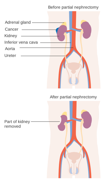
Before and after partial nephrectomy: the tumor is removed with a margin, and the rest of the kidney stays — and keeps working. Illustration: Cancer Research UK, CC BY-SA 4.0

If the kidney can’t be saved: radical nephrectomy
When radical nephrectomy is the right operation, I perform it robotically as well — single-port or multiport depending on the tumor’s size and anatomy. The operation removes the kidney with its surrounding fat, and the stay and recovery track closely with partial nephrectomy: one night in the hospital, and a similar arc back to normal life.
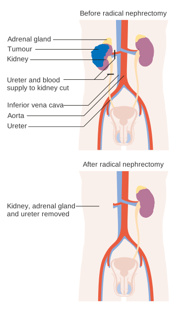
Before and after radical nephrectomy: the affected kidney is removed entirely, and the remaining kidney takes over. Illustration: Cancer Research UK, CC BY-SA 4.0

The reassuring truth about living with one kidney: a healthy solitary kidney compensates remarkably well. Most people notice no difference in daily life — no dialysis, no diet overhaul, no activity restrictions once healed. We monitor your kidney function with simple blood tests, and it typically stays stable for life.
Your hospital stay
Plan on one night. You’ll be walking and eating the evening of surgery — because the bowels are untouched, there’s no waiting for them to “wake up.” Most patients need little or no opioid medication. There’s no urinary catheter to go home with; occasionally, for complex reconstructions, I leave a small drain for a few days, removed at follow-up.
Recovery timeline
- Days 1–3: Home. Walking daily, stairs fine, most patients on over-the-counter pain medication only.
- Week 1: Back to desk work for most.
- Weeks 2–3: Energy returns to near-normal; driving and travel are comfortable.
- Weeks 4–6: Full activity, exercise, and lifting.
Risks, honestly
Partial and radical nephrectomy are safe operations in experienced hands, but you deserve the real list:
- Bleeding — the kidney is one of the most blood-rich organs in the body. Significant bleeding during surgery is uncommon; rarely, bleeding can occur days to weeks afterward and require a procedure to control.
- Urine leak (partial only) — the repaired collecting system can occasionally leak while healing, sometimes requiring a temporary internal stent.
- Loss of kidney function — some function in the operated kidney is always at stake; my technique choices are built around minimizing it.
- Conversion — rarely, a planned partial must become a radical nephrectomy for safety or cancer control. I’ll discuss this possibility with you beforehand, honestly.
- The standard surgical risks — infection, hernia at the incision, injury to nearby structures, anesthesia risks — all uncommon.
Follow-up after surgery
Pathology returns in about a week, and we review it together — the tumor type, grade, and margins. After that, follow-up is periodic imaging and kidney-function labs: more frequent in the first two years, then spacing out, with the schedule tailored to your tumor’s specific risk. Most patients with localized kidney cancer are followed for five or more years and never see it return.
Frequently asked questions
Do I need a biopsy before surgery? Usually not — this surprises people. Kidney imaging is accurate enough that most masses are diagnosed and treated based on the scan, and unlike the prostate, a suspicious kidney mass that needs removal is removed regardless of what a needle shows. Biopsy earns its place in specific situations — when surveillance is being considered, when the diagnosis is genuinely in doubt, or before ablation — and I’ll tell you honestly if yours is one.
How long does the surgery take? Typically two to three hours, depending on tumor complexity. You’ll be at the hospital most of the day.
Will I need dialysis? What is life with one kidney like? Dialysis is essentially never needed after kidney cancer surgery when the other kidney is healthy. Even after radical nephrectomy, the remaining kidney takes over and life continues without restrictions — the main change is that we protect it: sensible blood pressure control, hydration, and periodic labs.
When can I drive, work, fly, and lift? Driving: once you’re off pain medication and comfortable, usually within a week. Desk work: about a week. Flying: fine within a couple of weeks. Lifting and hard exercise: 4–6 weeks, to protect the incision while it strengthens.
What will my follow-up imaging look like? Periodic CT or ultrasound plus kidney-function labs — typically every six months at first, then annually, adjusted to your tumor’s risk. It’s a light schedule that mostly serves to confirm you stay well.
Considering this procedure?
I see patients at Swedish in Seattle and care for patients across Washington and the Pacific Northwest. Call to schedule a consultation.
Call (206) 386-6266